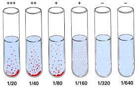
W.B.C.S. Examination Notes On – Widal Test Procedure – Medical Science Notes.
Widal test can be done in two ways
- Rapid slide test: It may be qualitative or quantitative and performed on slide.Continue Reading W.B.C.S. Examination Notes On – Widal Test Procedure – Medical Science Notes.
- Tube test: It is performed in tubes, is quantitative and requires incubation. It is considered more reliable than slide test.
SLIDE TEST
- Place one drop of positive control on one reaction circles of the slide.
- Pipette one drop of Isotonic saline on the next reaction cirlcle. (Negative Control).
- Pipette one drop of the patient serum to be tested onto the remaining four reaction circles.
- Add one drop of Widal TEST antigen suspension ‘H’ to the first two reaction circles. (PC & NC).
- Add one drop each of ‘O’, ‘H’, ‘AH’ and ‘BH’ antigens to the remaining four reaction circles.
- Mix contents of each circle uniformly over the entire circle with separate mixing sticks.
- Rock the slide, gently back and forth and observe for agglutination macroscopically within one minute.
SEMI-QUANTITATIVE METHOD
- Pipette one drop of isotonic saline into the first reaction circle and then place 5, 10, 20, 40, 80 ul of the test sample on the remaining circles.
- Add to each reaction circle, a drop of the antigen which showed agglutination with the test sample in the screening method.
- Using separate mixing sticks, mix the contents of each circle uniformly over the reaction circles.
- Rock the slide gently back and forth; observe for agglutination macroscopically within one minute.
STANDARD TUBE TEST METHOD
In Widal Test, two types of tubes were originally used:
(1) Dreyer’s tube (narrow tube with conical bottom) for H agglutination and
(2) Felix tube (short round-bottomed tube) for O agglutination.
Now a days 3 x 0.5 ml Kahn tubes are used for both types of agglutination.
- Take 4 sets of 8 Kahn tubes/test tubes and label them 1 to 8 for O, H, AH and BH antibody detection.
- Pipette into the tube No.1 of all sets 1.9 ml of isotonic saline.
- To each of the remaining tubes (2 to 8) add 1.0 ml of isotonic saline.
- To the tube No.1 tube in each row add 0.1 ml of the serum sample to be tested and mix well.
- Transfer 1.0 ml of the diluted serum from tube no.1 to tube no.2 and mix well.
- Transfer 1.0 ml of the diluted sample from tube no.2 to tube no.3 and mix well. Continue this serial dilution till tube no.7 in each set.
- Discard 1.0 ml of the diluted serum from tube No.7 of each set.
- Tube No.8 in all the sets, serves as a saline control. Now the dilution of the serum sample achieved in each set is as follows: Tube No. : 1 2 3 4 5 6 7 8 (control) Dilutions 1:20 1:40 1:80 1:160 1:320 1:640 1:1280.
- To all the tubes (1 to 8) of each set add one drop of the respective WIDALTEST antigen suspension (O, H, AH and BH) from the reagent vials and mix well.
- Cover the tubes and incubate at 37° C overnight (approximately 18 hours).
- Dislodge the sedimented button gently and observe for agglutination.
Positive: Agglutination within a minute
Negative: No agglutination indicative of absence of clinically significant levels of the corresponding antibody in the patient serum.
- The sample which shows the titre of 1:100 or more for O agglutinations and 1:200 or more for H agglutination should be considered as clinically significant (active infection).
- Demonstration of 4-fold rise between the two is diagnostic.
- H agglutination is more reliable than O agglutinin.
- Agglutinin starts appearing in serum by the end of 1st week with sharp rise in 2nd and 3rd week and the titre remains steady till 4th week after which it declines.
- Rising antibody titre is more convincing evidence of infection than positive tests alone. It is preferable to test two specimens of sera at an interval of 7 to 10 days to demonstrate a rising antibody titre.
- Low antibody titres are common in normal individuals and do not suggest an infection.
Please subscribe here to get all future updates on this post/page/category/website


 Toll Free 1800 572 9282
  mailus@wbcsmadeeasy.in


