

 +919674493673
  mailus@wbcsmadeeasy.in


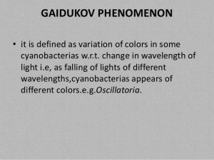
Botany Notes On – Gaidukov Phenomenon – For W.B.C.S. Examination.
Gaidukov phenomenon– this phenomenon occurs in algae. It is the phenomenon in which algae absorbs maximum light to perform photosynthesis.Continue Reading Botany Notes On – Gaidukov Phenomenon – For W.B.C.S. Examination.
In this process, the pigment composition changes to increase its efficiency for the maximum adoption of light. Gaidukov is also known as complementary chromatic adaptation.
The most clear-cut examples are to be found among the blue-green algae (Cyanobacteria), and it was indeed in a blue-green algal species that ontogenetic chromatic adaptation was first described. Gaidukov (1902) observed that Oscillatoria rubescens was red in colour when grown in green light, and blue-green when grown in orange light: he attributed these colour changes to the synthesis of different kinds of pigment. Boresch (1921) showed that the colour changes are due to shifts in the types of biliproteins synthesized by the algae: the red cells contain predominantly phycoerythrin, the blue-green cells mainly phycocyanin.
This phenomenon was considered by Engelmann and Gaidukov (1902) to be an example of complementary chromatic adaptation, the pigment induced by a specific waveband being one which absorbs that waveband (phycoer-ythrin and phycocyanin have their absorption peaks in the green (^565 nm) and the red (^620 nm), respectively): ‘complementary’ because the pigment has the complementary colour to that of the light that induces it.
Not all blue-green algal species show chromatic adaptation, and among those that do there are variations in the form that adaptation takes. Tandeau de Marsac (1977) separated cyanobacteria into three groups. In group I strains the biliprotein composition is not affected by the spectral quality of the light in which they are grown, i.e. they lack chromatic adaptation.
In group II strains, only the phycoerythrin content of the cells is affected by light quality, being very low in red light and high in green light; phycocyanin content remains high in cells grown under either kind of light. In group III strains, the synthesis of both the major biliproteins is affected by the spectral composition of the light: like the previous group their phycoerythrin content is high in green light and low in red, but their phycocyanin content, while still substantial in green light, is 1.6- to 3.7-fold higher in red light. Of 69 strains examined, 25 were in group I, 13 in group II and 31 in group m.176,1344
The phycocyanin formed in group I and group II strains has two polypeptide subunits, a and b. Bryant (1981) found that in 24 of the 31 group III strains, the phycocyanin formed in green light also contained just two subunits, but the phycocyanin from cells grown in red light contained four subunits, a1, a2, b1 and b2. The particular subunits present in phycocyanin in the green-light-grown cells were a2 and b1.
These, being always present, Bryant referred to as constitutive: the other two, a1 and b2, formed in red light, he referred to as inducible. Thus, in these 24 strains it appeared that the increased phycocyanin formation induced in red light consisted of synthesis of different kinds of phycocyanin subunit. It was not possible to determine from the data whether the new phycocyanin subunits associated to form a distinct phycocyanin species, (a1b2)n, or whether they aggregated with the two constitutive subunits, a2 and b1, to form a hybrid phycocyanin. In the remaining seven group III strains, no a1 and b2 phycocyanin subunits were found in red-light-grown cells: the extra phycocyanin in these cases may have consisted just of additional a2 and b1 subunits. In Tolypothrix tenuis, Ohki et al. (1985) found that chromatic adaptation in green light resulted in a one for one substitution of phycoerythrin for phycocyanin, so that phycobilisome size remained constant.
Ohki and Fujita (1992) found that cells of the marine cyanophyte Phormidium sp. C86 were coloured dark green when grown in red light, and purple-red when grown in green light, this change in colour being due to a massive increase in the cellular content of phycoerythrin. The bili-protein composition (molar basis) in red light was 13% phycoerythrin, 64% phycocyanin and 22% allophycocyanin. In green light this changed to 82%, 10% and 8%, respectively. The phycobilisomes in cells grown in green light were twice as large as those in red-light-grown cells.
Please subscribe here to get all future updates on this post/page/category/website