


 Toll Free 1800 572 9282
  mailus@wbcsmadeeasy.in


Life-Cycle – Pathogenecity And Control Of Leishmania – Zoology Notes – For W.B.C.S. Examination.
Leishmania donovani causes visceral leishmaniasis. The disease is also known as Dum-Dum fever, Asian fever, Assam fever or infantile splenomegaly in various parts of the world.Continue Reading Life-Cycle – Pathogenecity And Control Of Leishmania – Zoology Notes – For W.B.C.S. Examination.
- The parasite is named after its discoverers-Leishman and Donovan. Both Leishman and Dobovan reported the parasite simultaneously in the same year, 1903.
- Leishman demonstrated the parasite in the spleen smear of a soldier in England, who died of fever contracted at Dum-Dum in Kolkata.
- Donovan found the same in the spleen smear of a patient suffering from kala-azar in India.
- The san fly (Phlebotomus argentipes) was identified as a vector of the disease by Indian Kala-azar commission (1931-1934).
Habitat
- Leishmania donovani is an obligate intracellular parasite of man and other mammalian hosts.
- Promastigote forms of the parasite are found in sand fly and in culture
- Intracellular Amastigote forms are found in human in reticuloendothelial cells of the spleen, bone marrow, liver, intestinal mucosa and mesenteric lymph nodes.
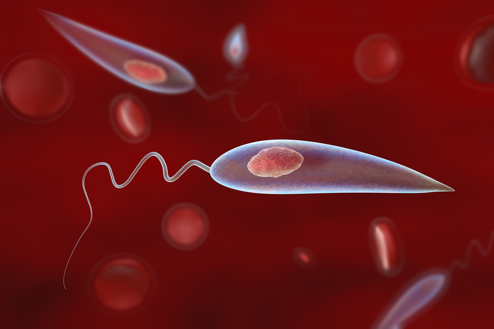
Morphology
- The parasite exists in two forms: Amastigote and Promastigote.
1. Amastigote
- Amastogote is the aflagellar stage of the parasite.
- The parasite at Amastigote stage are found in man and other mammalian hosts.
- They are found inside monocytes, polymorphonuclear leucocytes or endothelial cells.
- Amastigotes are small, round to oval bodies measuring 2-3 µm in length.
- They are also known as LD (Leishman Donovan) bodies.
- Cell membrane is delicate and can be demonstrated only in fresh specimen.
- The nucleus is less than 1 µm in diameter, oval or round and is usually situated in the middle of cell.
- A rod –shaped kinetoplast lies at the right angles to the nucleus. It comprises of DNA containing body and a mitochondrial structure.
- Axoneme (rhizoplast) arises from the kinetoplast and extends to margin of the body. It represent the foot of the flagellum.
- Vacuole, which is clear unstained space, lies alongside the axoneme.
- They are stained well with Giemsa or Wright stain.
- In a Giemsa stained preparation the cytoplasm surrounded by a limiting membrane appear pale blue. The nucleus relatively is larger and stained red. The kinetoplast stained deep red.
- Amastigote divides by binary fission at 37°C.
2. Promastigote
- Promastigotes are found in the digestive tract of sand fly (vector) and in the culture media.
- The fully developed promastigotes are long, slender and splindle-shaped. They measure 15 to 25 µm in length and 1.5 to 3.5 µm in breadth.
- A single nucleus is situated centrally.
- The kinetoplast lies transversely near the anterior end.
- The flagellum is single, delicate and measures 15- 28 µm and may be of same length as the body or even longer, projecting from front. The flagellum does not curve round the body of the parasite and therefore there is no undulating membrane.
- With Leishman stain, the cytoplasm appears blue, the nucleus pink or violet and the kinetoplast bright red.
- Promastigote multiplies by binary fission at 27°C.
Life cycle of Leishmania donovani
- The parasite has two stages in its life cycle:
- Amastigote form: occurring in humans and mammals.
- Promastigote form: occurring in sandfly.
- The parasite is transmitted to man or other vertebrate hosts by the bite of blood sucking female sand fly.
- During the blood meal, the sand fly deposits promastigote on the surface of the skin.
- These promastigotes are immediately phagocytized by macrophages and other types of mononuclear phagocytic cells. In these cells, promastigotes transform into the tissue stage of the parasite; amastigotes.
- The amastygotes multiply by simple binary fusion inside Reticuloendothelial system to form large number of amastigotes.
- The infected cell eventually becomes packed with the parasite.
- The host cell is thereby enlarged and ruptures.
- The parasites liberated infects new cells and the cycle is repeated and proceed to infect other mononuclear phagocytic cells.
- Some of the free amastigotes are phagocytosed by the neutrophils and macrophages in the blood stream.
- Free amastigotes are ingested by female sand fly during a blood meal from infected host.
- In the midgut of sand fly, the amastigotes are transformed within 72 hours through a series of flagellated intermediate promastigote forms to flagellated promastigotes.
- These promastigotes multiply by binary fusion and produce large numbers of promastigotes completely filling luman of the gut.
- After a period of 6-9 days, the promastigotes migrate from the midgut to the pharynx and buccal cavity of sand fly leading to heavy pharyngeal infection of the sand fly that feeds on plant juice after first blood meal.
- Bite of sandfly transmits the infection to new host and the life cycle is repeated.
Mode of transmission
- The infection is transmitted to Human mainly by the bite of vector sandfly of genus Phlebotomus and genus Lutzomyia.
- Less frequently the infection is transmitte by:
- Blood transfusion, congenitial infection, accidental inoculation of cultured promastigotes in the lab workers and sexual intercourse.
- Males are affected more due to increase exposure through the occupation and leisure activities.
Pathogenesis of Leishmania donovani
- After the inoculation of promastigotes by sand flies, they are deposited on the surface of skin and bind to macrophages in the skin.
- The sand fly, liberates biologically active substances, which promote infectivity of promastigotes by partially deactivating fixed macrophages in the skin.
- The outcome of leishmania infection appears to depend on the complex interaction between the parasite’s virulence and the immune response of the host.
- Promastigotes activate complement through the alternative pathway and are opsonized.
- They produce activated products of complement such as C3b or C3bi. These activated products bind with two specific receptors present on the outer membrane of promastigotes.
- The receptors are- a 63kD mol. wt glycoprotien (gp63) and a lipophosphoglycan (LPG). These receptors play an important role in the parasites- macrophage interaction.
- These receptors bind with complement receptors (CR3 and CR1) present on surface of macrophages either directly or through bound C3b or C3bi receptors.
- The most important immunological feature is a marked suppression of the CMI to leishmanial antigens. In persons with asymptomatic self-resolving infection, T-helper cells predominate, although immune suppression years later can result in disease. An overproduction of both specific Ig and non-specific Ig also occurs. The increase gamma globulin leads to reversal of the albumin-globulin ratio commonly associated with this disease.
- Leishmaniasis is a disease that involves the RE system. Parasitized macrophages disseminate the infection to all parts of body but more to the spleen, liver and bone marrow. The spleen is enlarged, with a thickening of the capsule and is soft and fragile, its vascular spaces are dilated and engorged with blood. The reticular cells are markedly increased and packed with the amastigote forms of the parasite. In the liver, the kupffer cells are increased in size and number and infected with amastigote forms. Bone marrow turns hyper plastic and parasitized macrophages replace the normal hemopoietic tissue.
- Proliferation and destruction of Reticuloendothelial cells of the internal organs and heavy parasitization of external organ by parasitized cells are the characteristic pathological changes seen in visceral leishmaniasis.
Please subscribe here to get all future updates on this post/page/category/website
Want to keep yourself updated about WBCS and other Govt. exams? Submit the form here-

For all WBCS Exam etc. course details CLICK HERE
Your information will *never* be shared or sold to a 3rd party.