

 Toll Free 1800 572 9282
  mailus@wbcsmadeeasy.in


W.B.C.S. Examination Notes On – Microsporogenesis – Botany Notes.
During the development of the microsporangium, the anther is seen at first as a homogeneous mass of meristematic cells, oblong in cross-section and surrounded by an epidermis.Continue Reading W.B.C.S. Examination Notes On – Microsporogenesis – Botany Notes.
It then becomes more or less four-lobed and four longitudinal rows of archesporial cells are differentiated. The archesporial cells are marked off from the surrounding cells by their more deeply staining cytoplasm and conspicuous nuclei.
There may be only one such archesporial cell in each of the four lobes as in Boerhaavia, etc., or there may be more of them forming a plate (Ophiopogon, etc.).
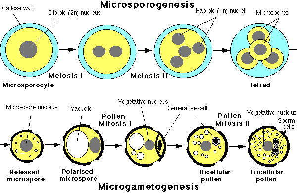
Longitudinally, also, there may be one to many of them. Each archesporial cell now cuts off a primary parietal cell towards the epidermis and a primary sporogenous cell on the inner side .
The parietal cell now divides by periclinal and anticlinal walls giving rise to several layers of cells forming the wall of the anther while the sporogenous cell usually divides a few times giving rise to a number of microspore or pollen mother cells .
The innermost layer of wall cells directly abutting on the sporogenous tissue forms the tapetum which is a nutritive tissue nourishing the developing microspores . The wall cells just below the epidermis form the endothecium which later loses the cell contents, usually becomes fibrous, and forms the dry coat of the mature anther in which the epidermis becomes rather inconspicuous.
Between the tapetum and the endothecium there are one to three middle layers of cells. The middle layers and the tapetum are usually crushed by the time actual meiosis occurs in the sporogenous cells.
During microsporogenesis (i.e., development of microspores or pollens) the nucleus of each microspore mother cell undergoes meiosis or reduction division ultimately giving rise to four haploid (i.e., possessing ‘n’ number of chromosomes) nuclei.
These four nuclei are arranged tetrahedrally and are soon invested with cell walls. Many variations are known of this typical pattern of meiosis, e.g., in maize a wall is formed across the dyad (2-nucleated condition).
They are now the microspores or pollens which soon dry up and become powdery while the tapetum becomes absorbed.
The anther now becomes a dry structure, the partition walls between the sporangia (i.e., loculi) are usually destroyed and the microspores (pollens) are soon liberated by dehiscence of the anther.
The tapetal cells often become multinucleate and play a great part in the nutrition of the pollens. Sometimes they develop a Plasmodium after disintegration and play a part in the development of the exine of the pollen. Even a part of the sporogenous tissue may break down and serve for nutrition instead of developing spores.
While the pollens are dry and powdery in most flowers, peculiar conditions are often let with. In Annona, Elodea, Typha, etc., the four spores in a tetrad never separate but form compound pollen grains.
In the Mimoseae 8 to 64 pollens often aggregate together while in the gynostegium of Calotropis and the gynostemium of orchids all the pollens of each anther lobe from a characteristic mass called pollinium. Each pollinium is provided with a stalk called caudicle and a sticky base called disc or corpusculum.
The pollen or the microspore is a very minute structure (0.025 to 0.125 mm in diameter). It is unicellular and usually round although it may be oval, pyramidal, polyhedral, etc. It is provided with two coats—an inner, delicate cellulose layer called intine and an outer tough, cutinised layer called exine or extine. The exine is often sculptured or provided with spines, warts, etc. Occasionally, it is smooth.
The exine may have a waxy coating to render the pollen more or less waterproof. Very often, there are some definitely thinner, circular spots or slits in the exine called germ pores or slits.
These weak spots are utilised during the germination of the pollen. The pores are sometimes provided with lids which open out like valves during germination. Very often, before the pollen is discharged from the anther it becomes binucleate , the original single nucleus dividing-into a tube nucleus and a generative nucleus.
The latter, with some cytoplasm surrounding it, becomes the generative cell (described later). Sometimes the pollen may even become trinucleate (Fig. 410), as in most cereal crops, by complete development of the male gametophyte even before it is shed.
Please subscribe here to get all future updates on this post/page/category/website